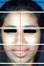
- Upper part or the forehead: From hairline to glabella.
- Middle part or the midface: From glabella to subnasale.
- Lower part or the lower face: From subnasale to menton.
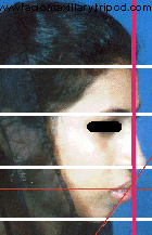
In the side view (profile), the face can be convex, concave or straight type. To evaluate the horizontal relations of the soft tissues a line can be dropped perpendicular from the Frankfort horizontal plane through the root of the columella (subnasale). This perpendicular line passes through the upper vermilion border, 2 mm out to the lower vermilion border and 4mm (+/- 2 mm) anterior to the menton.
Some salient features that tell about the harmony and balance of the face are:
- Middle third is equal to Lower third of the face.
- Subnasale to stomion is half that of stomion to menton.
- Subnasale to lower lip vermilion border is equal to lower lip vermilion border to menton.
- Interlabial distance is 0 - 3mm at rest.
- The upper lip margin lies on the gingival margin while smiling.
- Width of the nose is equal to or few mm wider than the inner intercanthal distance.
- Position of the midline of the chin gives the face its symmetry.
- Normal naso-labial angle ranges between 900 - 1100.
- Labiomental fold gives a pleasant definition to the face.
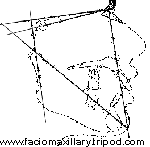 Lateral Cephalogram is traced to analyze the proportion
of the various parts of the face. This will help to find out the areas of
disproportion by marking out the various points, but should be correlated with
clinical observations. Innumerable analyses have been proposed but great
variations are seen in the "normal values" between different human races. More
importance should be given for soft tissues measurements than skeletal
analysis. The points, lines and angles marked are very arbitrary and different
values can be obtained from different analysis of the same person. COGS
analysis is the most commonly used and it describes the horizontal and
vertical position of the facial bones by use of a constant coordinate
system. Both the skeletal and soft tissue evaluation can be done by this
analysis.
Lateral Cephalogram is traced to analyze the proportion
of the various parts of the face. This will help to find out the areas of
disproportion by marking out the various points, but should be correlated with
clinical observations. Innumerable analyses have been proposed but great
variations are seen in the "normal values" between different human races. More
importance should be given for soft tissues measurements than skeletal
analysis. The points, lines and angles marked are very arbitrary and different
values can be obtained from different analysis of the same person. COGS
analysis is the most commonly used and it describes the horizontal and
vertical position of the facial bones by use of a constant coordinate
system. Both the skeletal and soft tissue evaluation can be done by this
analysis.
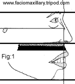
Fig:1 – Pre operative profile tracing with vertical excess of maxilla marked.
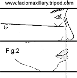
Fig:2 – Facial profile tracing after Le-Fort I osteotomy.
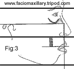
Fig:3 – Anterior maxillary osteotomy traced.
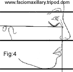
Fig:4 -- Profile tracing of the final surgical outcome.
A cut and paste technique on the tracing paper is used for predicting the outcome.
Computer prediction is now available due to the development of various software programs. This helps in visualizing the postoperative results better, but chances are, expectations become higher and the surgeon may have difficulty in producing the same results clinically.
- Get an idea about the extent of bone reduction required in the surgery.
- Understand the post-operative relations of the jaws.
- Understand the post-operative occlusion.
- Helps in the fabrication of splints.
- Decide about the post-surgical orthodontic treatment.
Wafer thin occlusal splint should be fabricated to prevent the condyle from being unseated vertically, which can cause "condylar sag" post-surgically. The splint should allow the teeth to occlude properly in the preplanned occlusion.
