Surgical Procedure:
Steps 5 to 8
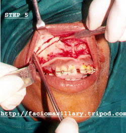
Measurements made to assess the amount of bone to be removed during the presurgical analysis is marked. Lateral wall of the maxilla is cut with the help of a micromotor using a 590 straight fissure surgical bur. Copious irrigation is to be maintained with saline. In most cases the cut is kept parallel to the occlusal plane and extends up to the pterygoid.
Use a thin small osteotome and tap gently and carefully to fracture the medial (lateral nasal wall) and posterior wall of maxilla. The nasal mucosa is protected from injury with a periosteal elevator while completing the osteotomy.
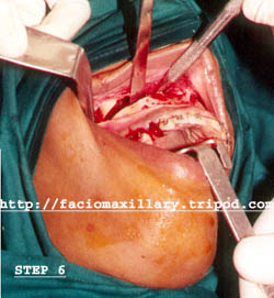
The pterygoid plate is separated from the maxilla using a pterygoid osteotome. The pterygoid hamulus is felt and palpated with the ball of the index finger to prevent excess in chiseling and damage to the soft tissues. Failure to separate the tuberosity from the pterygoid plates will cause difficulty in downfracture or an unfavorable fracture.
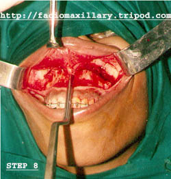
Repeat the steps 5 through 7 on the other side. Make sure that the cuts are at the same level as on the initial side.
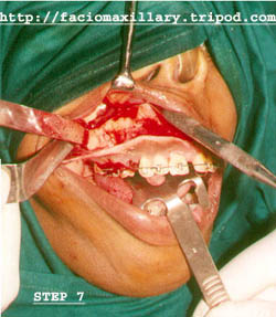
The nasal septum cartilage and vomer is separated from the maxilla using a septal gouge or osteotome. After the fracture of the anterior nasal spine angle the gouge towards the floor of the nose.